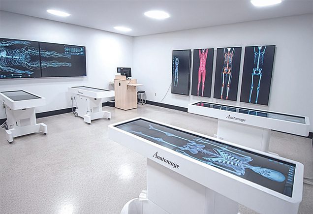
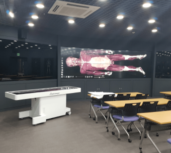
Anatomage Table
The World’s First and Only Real-Tissue Virtual Dissection Table
Advanced 3D anatomy
visualization and virtual
dissection tool for
learning anatomy and
physiology.
The Anatomage Table is the Real-Tissue based & technologically most advanced 3D anatomy visualization system for anatomy and physiology education The operating table form factor combined with Anatomage’s renowned radiology software апа clinical content separates the Anatomage Table from any other imaging system on the market. The team traces out anatornical structures,
including blood vessels апа nerves, оп each 2D slice. The slices are then stacked
tо recreate segmented 3D anatomy. Because over 2,500 anatomical structures
are segmented, each structure can be viewed or virtually dissected individually or in context with one another.
It is designed to support the most challenging tasks in healthcare education.
What’s New in Anatomage Table 12

Anatomage Table 12 introduces enhanced anatomy visualisation, expanded clinical learning content, and advanced functional physiology simulations. Built on real human cadaver data, it enables students and healthcare professionals to explore anatomy, pathology, radiology, and clinical procedures with exceptional accuracy.
The latest platform also features new pathological specimens, the Precision Anatomy Model, Multi Body comparisons, and improved educational tools that support immersive, interactive, and clinically relevant learning experiences.
- Explore a complete real human cadaver dataset featuring. metastatic pancreatic cancer
- Examine pathological anatomy across multiple organ systems with exceptional anatomical detail.
- Visualise disease progression and anatomical changes through high-resolution 3D anatomy.
- Study clinically relevant pathology integrated with anatomy, radiology, and physiology
- Ideal for pathology, oncology, anatomy, and advanced medical education programs.
- An 84-year-old female with pancreatic cancer, featuring healthy anatomy side-by-side with localised tumour metastases

- The Anatomage Table 12 combines thousands of real human anatomy and pathology cases with advanced 3D visualization technology, delivering the world’s most comprehensive virtual dissection experience for anatomy and clinical education.
- This advanced simulator offers extreme visual clarity into the various stages of labor, including the cervical
dilation, infant rotation and head movements, and release of the placenta. - Interactive interface
- Adjustable speed
- Ideal for obstetrics, maternal education ап care
- Medical professionals ап students can remove anatomical parts to explore the anatomical transformations
occurring throughout the birthing process.

- The upgraded Cardiology simulation is a collection оf arrhythmias, enabling students to compare a normal heartbeat with irregular rhythms such as atrial fibrillation, ventricular fibrillation, and various heart blocks.
- Cardiac activity can be tracked on an ECG
- A-Fib, V-Fib, and 1st, 2nd, and 3rd degree heart blocks
- Ideal ог cardiovascular education and training

- As part of the Blood Flow tool, the Vascular Grow feature maps out the blood vessels in the cadaver, giving medical students a thorough view of how the heart and blood vessels are connected.
- Doctors can use this tool to see where important arteries split and veins come together. It’s also helpful for explaining to heart patients how their blood vessels work and where to place catheters safely.
- Interactive view of the blood flow
- Visualise arterial bifurcation and venous convergence
- Identify vascular anomalies or conditions
Point-to-Point Dissection
- A step-by-step procedure that helps students easily visualise anatomical structures and diseases.
- Students initiate the process by marking an entry point, then proceed to dissect through a connected point.
- Provides a comprehensive perspective for examining tumors from multiple angles.
- Explore the spatial relationships
- Practice customized surgical procedures
- Ideal for surgical planning, sport injuries inspection

Ultrasound Viewer
- The Ultrasound Fan view, revealing the internal structures
- An ideal resource for sonography training
- Learn the impact of probe rotation and angle on the resulting images.
- Real-time, cross-sectional images
- Ideal for sonography, radiology
- Suitable for Education and professional settings

CLINICAL-BASED LEARNING TOOLS
CLINICAL-BASED LEARNING TOOLS
- Personalized navigation
- Ideal for group sessions, autonomous learning
- This tool enable students to quickly identify anatomical structures and distinguish adjacent structures through various colors.


Trusted by Top Organisations


Unique Simulation Features
in-situ and 'Just in time' training
- 6 Full-Body Real Human Cadavers for comprehensive virtual dissection and anatomical exploration.
- World’s first life-size virtual dissection table that transforms your anatomy lab into a high-tech workstation where students can acquire real-world clinical experience.
- No chemicals , no unpleasant smells, no regulations and a higher student adoption rate over traditional cadavers.
- 6 gross anatomy cases, 40 high resolution regional anatomy cases, and more than 1,600 pathological examples, including animal cases. These are high resolution and high quality cases are unique to Anatomage.
Advantages of Anatomage Table
Based education is proven to be more effective. Growing publications show improved test scores, more efficient class and lab sessions, and student acceptance.
The Table allows students to interact with young and well preserved digital cadavers instead of aged and degenerated bodies. Students are also exposed to different anatomical variations and a large number of pathological variations.

What Makes Anatomage Table Unique?
The table has a collection of thousands of real human cases and digitized them in highest possible resolution and spent years painstakingly separating individual structure from each slice.
Our innovative simulation products provide immersive training experiences that enhance clinical proficiency and decision-making. Get in touch with us to discover how our solutions can elevate your training and improve patient outcomes.”
Frequently Asked Questions (FAQs)
1. What is the Anatomage Table?
The Anatomage Table the world’s 1st life-size , Real -tissue dissected digital anatomy platform that enables virtual dissection, anatomy visualization, and medical education using real human cadaver data.
2. What is new in Anatomage Table 12?
Anatomage Table 12 introduces the Precision Anatomy Model, Multi-Body Comparison, a metastatic pancreatic cancer cadaver, enhanced pathology content, and improved learning tools.
3. Is the Anatomage Table based on real human cadavers?
Yes. Anatomage converts real human cadavers into highly detailed digital datasets, providing anatomically accurate and interactive learning experiences.
4. How many cadavers are included in Anatomage Table 12?
Anatomage Table 12 features 6 full-body real human cadaver datasets, including a metastatic pancreatic cancer specimen for pathology and oncology education.
5. What is the difference between traditional cadaver dissection and the Anatomage Table?
The Anatomage Table allows unlimited virtual dissections, eliminates chemical exposure, preserves anatomical structures, and provides integrated radiology and pathology learning.
6. Which courses can use the Anatomage Table?
The Anatomage Table is suitable for MBBS, BDS, Nursing, Physiotherapy, Allied Health Sciences, Radiology, Anatomy, and Medical Simulation programs.
7. Does Anatomage Table support CT, MRI, and X-ray imaging?
Yes. Anatomage integrates CT, MRI, X-ray, ultrasound, and other medical imaging modalities to enhance anatomical understanding and clinical correlation.
8. Can the Anatomage Table be used for pathology and oncology education?
Yes. Students can study pathological specimens, disease progression, anatomical abnormalities, and oncology cases using real patient-derived datasets.
9. Why do medical colleges choose Anatomage Table?
Medical institutions choose Anatomage Table for its real human anatomy visualization, virtual dissection capabilities, pathology content, radiology integration, and collaborative learning environment.
10. Where can I buy Anatomage Table in India?
Maverick Simulation Solutions is an authorized partner of Anatomage Inc. in India, providing product demonstrations, consultation, installation, training, and after-sales support.
11. Does Anatomage Table 12 include pathological cadaver models?
Yes. Anatomage Table 12 includes a Metastatic Cancer Cadaver, an 84-year-old female with metastatic pancreatic cancer, allowing learners to explore pathological anatomy, oncology, and disease progression through interactive 3D visualization based on real human cadaver data.